What is Congenital Heart Disease?
Quick Facts
- Congenital heart disease (CHD) develops when the heart is formed in the first two months of pregnancy.
- There are many categories of CHD with effects ranging from mild to severe.
- Most of these heart defects are treatable, and most people with CHD live into adulthood.
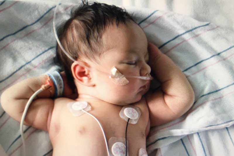
Sloan was born with tetralogy of Fallot.
Congenital heart disease (CHD) develops when the heart’s early formation is disrupted within the first two months of pregnancy. There are many causes for childhood heart problems, including defects from faulty embryo development, genetic causes and heart rhythm disturbances. In many cases, the exact cause can’t be identified. These cardiac defects can be diagnosed before birth or early in childhood.
“Congenital” means existing at birth. While “defect” describes a structural difference, many doctors and researchers use “disease” to reflect the full range of experiences people may have with CHD. There are several categories of possible childhood heart problems:
- Septal defects
- Conotruncal anomalies
- Right/left outflow tract obstruction
- Anomalies of the great arteries and veins
- Single ventricle physiology
- Coronary anomalies
- Valve abnormalities
- Complex cyanotic CHD
Heart defects can range from mild to severe. Some barely affect blood flow and cause no symptoms, while others significantly interfere with how blood circulates through the body.
Depending on the type of CHD, long-term complications can include:
- Pulmonary hypertension
- Systemic hypertension
- Arrhythmias
- Infective endocarditis
- Heart failure
- Kidney and liver disease
- Valve dysfunction
- Aortic dilation
- Thromboembolism
- Pregnancy-associated risks
Glossary of Congenital Defects and Common Types
Healthy Heart Function
A normal heart has valves, arteries and chambers that carry the blood in a circulatory pattern: body → heart → lungs → heart → body. When the heart works as it should, blood goes to the lungs for oxygen and then back out to the body. If valves, chambers, arteries or veins don’t develop normally, this blood flow can be disrupted. Congenital heart defects are malformations present at birth. They may or may not affect circulation.
Learn how a healthy heart works.
Aortic Valve Stenosis (AVS)
Aortic valve stenosis is a problem with the valve that carries blood from the heart to the body. The valve does not open fully and may also leak blood. When the blood flow out from the heart is blocked by a stiff valve, pressure builds up inside the heart and causes damage.
More information about aortic valve stenosis
Atrial Septal Defect (ASD)
An atrial septal defect is a hole in the wall that separates the upper chambers of the heart. This opening allows oxygen-rich blood from the left atrium to flow into the right atrium, where oxygen-poor blood is normally found. As a result, extra blood flows to the right side of the heart and to the lungs.
More information about atrial septal defect
Coarctation of the Aorta (CoA)
Coarctation of the aorta is a narrowing of the major artery (the aorta) that carries blood to the body. This narrowing reduces blood flow to different parts of the body and can make the heart work harder. CoA can cause high blood pressure or heart damage.
More information about coarctation of the aorta
Complete Atrioventricular Canal Defect (CAVC)
A CAVC defect is a large opening in the center of the heart that affects all four chambers. Because the walls and valves that normally separate the upper and lower chambers are not fully formed, oxygen-rich and oxygen-poor blood can mix. This makes the heart work harder and prevents blood from being routed properly to the lungs and the rest of the body.
More information about complete atrioventricular canal defect (CAVC)
d-Transposition of the Great Arteries
In d-transposition of the great arteries, the two major arteries are connected to the wrong heart chambers. The aorta, which should carry oxygen-rich blood to the body, instead arises from the right ventricle. The pulmonary artery, which should carry oxygen-poor blood to the lungs, arises from the left ventricle.
Because of this reversal, the blood flows in two separate loops instead of one continuous cycle:
- Body → heart → body (without going to the lungs for oxygen) or
- Lungs → heart → lungs (without delivering oxygen to the body)
To survive, a baby needs at least one natural “mixing” site — such as an atrial septal defect, a ventricular septal defect or a patent ductus arteriosus — so some oxygen-rich blood can reach the body. Doctors may create or enlarge a connection between the upper chambers of the heart using a balloon atrial septostomy to improve mixing.
Corrective surgery is usually required soon after birth to restore normal circulation.
More information about d-transposition of the great arteries
Ebstein’s Anomaly
Ebstein’s anomaly is a rare heart defect in which the tricuspid valve — the valve between the right atrium (upper chamber) and right ventricle (lower chamber) — is abnormally formed and positioned. Because the valve doesn’t close properly, blood can leak backward from the right ventricle to the right atrium. Many people with Ebstein’s anomaly also have an atrial septal defect (ASD), which allows blood to flow between the two upper chambers (atria) of the heart.
More information about Ebstein's anomaly
L-Transposition of the Great Arteries
In L-transposition of the great arteries, the heart’s lower chambers (the ventricles) and the two main arteries are switched. The right and left ventricles are reversed in position, and the aorta and pulmonary artery arise from the “wrong” ventricles.
Because both the ventricles and the arteries are switched, the blood still follows through a normal pathway:
- Oxygen-poor blood eventually reaches the lungs and
- Oxygen-rich blood is still sent out to the body
This is why L-TGA is often called a “double reversal” and is usually less immediately dangerous than d-TGA.
However, the ventricle that is normally designed to pump blood to the lungs must instead pump to the body over many years, which can lead to heart-function problems or valve issues later in life.
More information about L-transposition of the great arteries
Patent Ductus Arteriosus (PDA)
Patent ductus arteriosus is a congenital heart condition in which a temporary blood vessel (the ductus arteriosus) that normally closes shortly after birth remains open, allowing abnormal blood flow between the aorta and the pulmonary artery.
More information about patent ductus arteriosus
Pulmonary Valve Stenosis
Pulmonary valve stenosis occurs when the pulmonary valve is thickened or fused, preventing it from opening fully. This makes it harder for blood to flow from the right ventricle (lower chamber) into the pulmonary artery and on to the lungs. The heart must work harder to push blood through the narrowed valve.
More information about pulmonary valve stenosis
Single Ventricle Defects
Single ventricle defects are rare heart lesions in which one of the heart’s lower chambers (ventricles) is underdeveloped or cannot function normally. The affected ventricle may be too small, poorly formed or missing a working valve. As a result, the heart cannot pump blood effectively.
Hypoplastic Left Heart Syndrome (HLHS) — In HLHS, the structures on the left side of the heart, including the left ventricle, aorta and aortic valve, are severely underdeveloped. Openings in the heart and major vessels that normally close after birth may remain open so that blood can continue to flow to the body and lungs. HLHS requires urgent treatment soon after birth.
Pulmonary Atresia/Intact Ventricular Septum — In this defect, the connection from the right ventricle to the pulmonary artery does not form. Because the pulmonary valve and the opening beneath it are completely closed, blood cannot leave the right ventricle to reach the lungs. Instead, the body depends on other temporary pathways, such as the ductus arteriosus, which normally closes after birth to deliver blood to the lungs for oxygen.
Tricuspid Atresia — Tricuspid atresia occurs when the tricuspid valve and the opening from the right atrium to the right ventricle do not form. Blood from the body cannot flow normally into the right ventricle. Instead, it passes from the right atrium to the left atrium through an atrial septal defect, then to the left ventricle and out to the body — without being oxygenated. To receive oxygen, blood must find another pathway to reach the lungs, such as a ventricular septal defect (VSD) or a patent ductus arteriosus (PDA).
More information about single ventricle defects
Tetralogy of Fallot
Tetralogy of Fallot is a heart defect that includes four related problems:
- Ventricular septal defect (VSD): A hole between the lower chambers (ventricles) of the heart.
- Pulmonary stenosis: An obstruction that makes it harder for blood to flow from the right ventricle to the lungs.
- Overriding aorta: The aorta, which carries blood to the body, is positioned over the hole between the ventricles instead of over the left ventricle.
- Right ventricular hypertrophy: The muscle of the right ventricle becomes thickened because it must work harder to push blood through the obstruction to the lungs.
These four problems together reduce blood flow to the lungs and allow oxygen-poor blood to flow out to the body. This can cause cyanosis (“blue baby” appearance) if not treated.
More information about tetralogy of Fallot
Total Anomalous Pulmonary Venous Connection (TAPVC)
TAPVC is a heart defect in which the veins carrying oxygen-rich blood from the lungs do not connect to the left atrium as they should. Instead, these veins connect to other veins or to the right side of the heart.
Because of this abnormal connection, oxygen-rich blood mixes with oxygen-poor blood and does not flow normally from the lungs to the heart and out to the body. A hole between the upper chambers of the heart is needed so oxygenated blood can reach the body.
More information about total anomalous pulmonary venous connection (TAPVC)
Truncus Arteriosus
Truncus arteriosus is a heart defect in which one large blood vessel comes out of the heart instead of two separate vessels — one to the lungs and one to the body. Because of this, oxygen-rich and oxygen-poor blood mix together before leaving the heart. The body does not get enough oxygen-rich blood.
More information about truncus arteriosus
Ventricular Septal Defect (VSD)
A VSD is a hole in the wall (septum) that separates the heart’s two lower chambers, called the ventricles. Normally, this wall closes before birth, so oxygen-rich and oxygen-poor blood stay separate. When the opening remains, blood can flow between the ventricles, which may cause the heart to work harder, increase pressure in the heart and lungs or reduce the amount of oxygen delivered to the body.


