Testing for Heart Valve Problems
Echocardiogram
What is an echocardiogram?
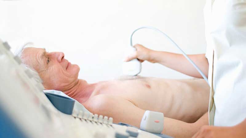
An echocardiogram (echo) is a test that uses high-frequency sound waves (ultrasound) to make pictures of your heart. The test is also called echocardiography.
Why do I need an echocardiogram?
An echocardiogram looks at your heart’s structure and checks how well your heart functions. The test provides information about:
- The size and shape of your heart.
- The size, thickness and movement of your heart’s walls.
- How your heart moves.
- The heart’s pumping strength.
- If the heart valves are working correctly.
- If blood is leaking backward through your heart valves (regurgitation).
- If the heart valves are too narrow (stenosis).
- If there is a tumor or other abnormalities around your heart valves.
Understanding Your Echo Results (PDF)(link opens in new window)
Measuring the valve gradient
The valve gradient is the difference in pressure on each side of the valve. When a valve is narrowed (a condition called stenosis), the pressure on the front of the valve builds up as blood is forced through the narrow opening. This causes a larger pressure difference between the front and back of the valve. The valve gradient can be used to determine the severity of the valve disorder.
- A leaking or regurgitating valve can also affect the pressure in both the heart chambers as well as surrounding blood vessels.
Measuring the valve area
The valve area is the size of the open valve. This is done using measurements during an echocardiogram. The valve area is in square centimeters and can be used to determine the severity of the valve disorder.
Measuring ejection fraction
- The ejection fraction describes how much blood the left ventricle pumps out with each contraction.
- A normal left ventricular ejection fraction is 50% to 70%. This means that between 50% and 70% of the blood in the left ventricle is pumped out with each heartbeat. When that number falls, especially below 40%, it can indicate a significant problem with the heart muscle. Learn more about ejection fraction.
Exercise stress test
Why is an exercise test sometimes needed to assess a valve problem?
Also called a stress test or treadmill test, exercise testing can provide valuable information in people with valvular heart disease, especially in those whose symptoms may be difficult to assess.
Your health care team uses an exercise test to provide additional information in asymptomatic people or people who have a history of symptoms. It also may be used in combination with echocardiographs, ECG, chest x-ray and cardiac catheterization.
Exercise testing helps evaluate changes in blood pressure and symptoms and the heart’s response to a more challenging workload. Because heart valve symptoms can develop slowly, you may not realize you have limited activity over time or attributed the change to “normal aging.” If you have a heart murmur, your health care team will note any changes in the murmur that happen during exercise. Read more about exercise testing.
Chest X-ray
What’s a chest X-ray?
A chest X-ray is a picture of the heart, lungs and bones of the chest. A chest X-ray can show the heart’s overall shape, but it can't show the inside structures of the heart.
Why is it done on someone with a valve condition?
A chest X-ray shows the location, size and shape of the heart, lungs and blood vessels. This can provide clues to a valve problem that include an enlarged or thickened heart and calcium deposits on the aorta or pericardium. Read more about chest x-ray procedures.
CT Scan
What’s a CT Scan?
The CT scan captures multiple X-ray images to create a cross-sectional image of the heart and lungs. Like an MRI, this test sometimes takes clearer pictures. Unlike MRI imaging, the CT scan uses the lowest radiation dose possible ‒ about as much as an X-ray.
Although an echocardiogram, including transesophageal echocardiography and transthoracic echocardiography is now the standard tool for assessing valvular heart disease, there are times when its effectiveness is limited in some people. A CT scan creates images of the valve anatomy and allows for evaluation of the severity of stenosis and regurgitation. A CT scan also can determine whether there are valvular lesions or nearby tumors affecting the function. Read more about CT scans.
Cardiac catheterization
How does cardiac catheterization help diagnose valve disease?
Although cardiac catheterization is most often used to look at the blood flowing to the heart muscle, it also can provide important information about narrowed heart valves, leaky heart valves or blood that is not flowing through the heart as it should.

View an illustration of cardiac catheterization.
For people with valve disease, a cardiac catheterization can:
- Measure blood pressure within the heart and oxygen in the blood
- Evaluate heart muscle function for moving blood through each chamber
- Help determine the best course of treatment
Cardiac catheterization is a minimally invasive procedure and isn’t necessary for every person who has a cardiac murmur or valve problem, but it can provide additional information when other tests may be inconclusive. Read more about cardiac catheterization.
Cardiac magnetic resonance imaging
Cardiac magnetic resonance imaging (CMR), also called cardiac MRI, is a painless, non-invasive imaging test used to assess the function and structure of the heart. It uses radio waves, magnets and a computer to create detailed pictures of your heart.
CMR has become an optimal technique in assessing people with heart valve disease without the need for radiation. Its enhanced diagnostic power can:
- Determine the type and severity of valve disease.
- Measure heart function or how much blood the left ventricle can pump out to the body.
People with any type of metal device inside the body should not have a CMR unless the device is certified as MRI safe.
Check with your doctor about the safety of CMR if you:
- Have a stent or artificial heart valve, or if you have had open-heart surgery recently.
- Are pregnant, especially during the first three months.
- Have tattoos or permanent (tattooed) makeup. You might feel some mild discomfort or a burning feeling on your skin from the metal in the darker inks of the tattoo.
- Have been told you have kidney problems.
Read more about magnetic resonance imaging.


