Family history and ultrasound help save woman with aortic dissection
By Deborah Lynn Blumberg, American Heart Association News


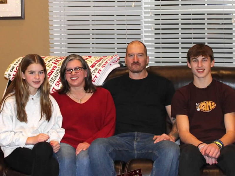
At her New Jersey home one summer evening, Sara Adair had just sent a text when a sharp pain shot through her chest. As the pain crept into her neck, a single thought seized her mind: Here it is.
A decade earlier, her dad, Sam Wisniewski, survived a tear in the wall of his aorta, the largest artery that carries oxygen-rich blood from the heart to the rest of the body. He was lucky: Up to 50% of people with aortic dissections die because they don’t get to the hospital in time, and tears can be difficult to diagnose.
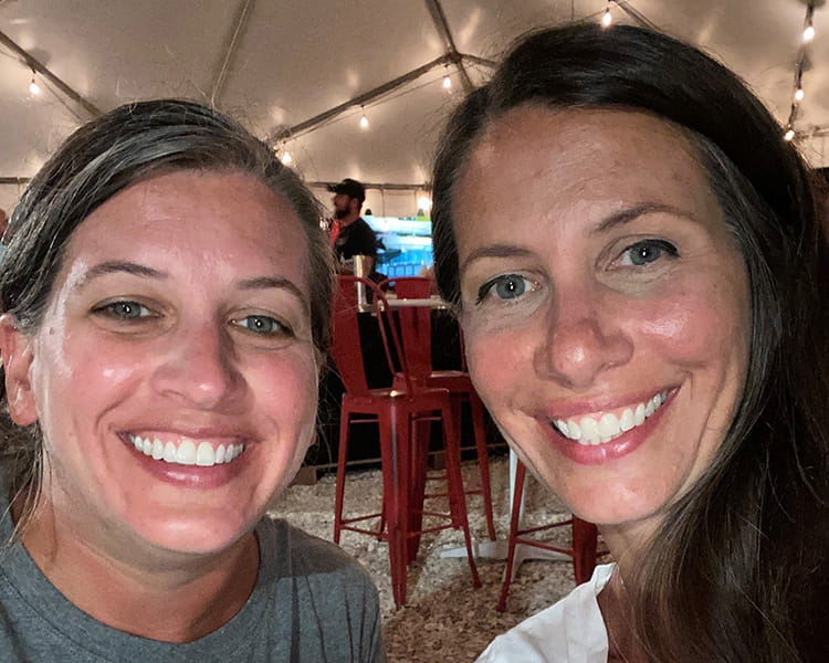
In 2022, just two years before Sara’s experience, her older sister, Dyan Kearns, survived a tear in the wall of her aorta. Dyan was then found to have Loeys-Dietz syndrome, a genetic connective tissue disorder that increases the risk of serious vascular issues like tears.
Tests showed Sara had the syndrome, too. She then saw a cardiologist and learned her aortic arch was slightly enlarged but still within normal limits. Her doctor recommended regular ultrasounds and CT scans, but no other changes. Still, Sara became even more vigilant about eating well. She also got back into running, although she knew she may not be able to out-diet and out-exercise her family health history.
Sara knew the aortic arch might become more enlarged, perhaps eventually requiring surgery. Her bigger fear was that her aorta might tear. And if it happened, when?
Chest pain turns into a crisis
Then came that summer evening when she felt the sharp pain in her chest and neck.
Before Sara could tell her husband, Brett, to call for help, she collapsed onto the living room floor. Seconds later, he found her face-down and called 911. Their son, Aaron, then 14, helped flip her over. Her lips were turning blue.
As first responders wheeled her away, Sara wanted to tell them about her dad and sister. She couldn’t. But she did manage to convey that she wanted to go to Robert Wood Johnson University Hospital, her workplace for 25 years. She’d been an X-ray technician, then an analyst in the radiology department. The hospital felt like home, and the staff felt like family.
An ultrasound tool helps save her life
In the ambulance, tests showed Sara was having a heart attack. Paramedics alerted the emergency room. Plans were made for her to undergo a cardiac catheterization, a standard way of unblocking her presumably clogged arteries.
Before taking Sara to the cath lab, a cardiology fellow examined her. Sara still couldn’t talk, so she was unable to tell the doctor about her family history and the possibility that she may have a torn aorta.
It was just a coincidence the doctor’s examination included using a tool he’d recently been trained on: a point-of-care ultrasound that gave him a peek inside her heart.
That’s how he determined Sara wasn’t having a heart attack. Her aortic wall had torn. It was flapping back and forth through her aortic valve. Blood was leaking back into her heart chambers.
Within minutes, the doctor canceled the cath lab and arranged for emergency surgery.
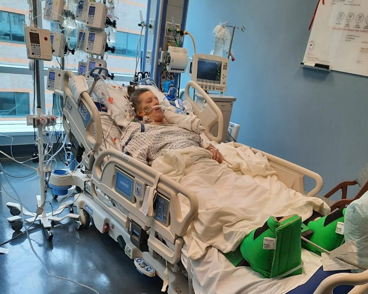
The torn – or dissected – part of Sara’s aorta and the leaking valve were replaced. However, during surgery, she had a stroke.
Complications mounted during her recovery in the hospital. She developed fluid in her chest, had three blood clots form in her left arm, and then learned she was allergic to the clot-busting medication.
Family life after surgery and stroke
Finally, after three weeks, Sara was well enough to go home – just in time for Aaron’s high school football season. Sara didn’t miss a single game.
The stroke left Sara with swelling and weakness in her left hand, plus blurry vision that gives her painful headaches. Each symptom has improved over time, and she hopes one day they’ll resolve. “It’s hard,” she said, “but livable.”
She goes to cardiac rehabilitation twice a week. Baseball, softball and rugby seasons keep her busy attending games to cheer on Aaron, now 15, and her daughter, April, 13. “But I’m also enjoying not doing things,” Sara said.
Meanwhile, Sara’s kids and nieces have seen a pediatric cardiologist and had cardiac ultrasounds. Genetic testing will determine if they also have Loeys-Dietz.
Sara often thinks about her care team’s quick response, especially when, at work, she bumps into a nurse or doctor who cared for her. She’s grateful for her positive outcome.
“That’s the essence of using the right technology at the right time,” said Dr. Partho P. Sengupta, chief of cardiology at Robert Wood Johnson University Hospital, an RWJBarnabas Health facility and Rutgers Health. He’s trained all of his cardiac fellows on the device. “There are two different pathways for heart attack and dissection, and minutes count.”
In February, Sara and her family reunited with her care team, including the EMTs. Sara thanked each person.
“If I had been treated as a heart attack, honestly I don’t think I would be here today,” Sara said. “The cardiology resident who was trained to use that ultrasound saved my life.”
Take charge of your heart health
Knowing her family history helped Sara recognize that something was seriously wrong. Go Red for Women, an American Heart Association initiative, helps women take charge of their heart health with information, resources and support to help prevent and manage cardiovascular disease.
Stories From the Heart chronicles the inspiring journeys of heart disease and stroke survivors, caregivers and advocates.