
 American Heart Association
 URGENT: 4X Match Extended
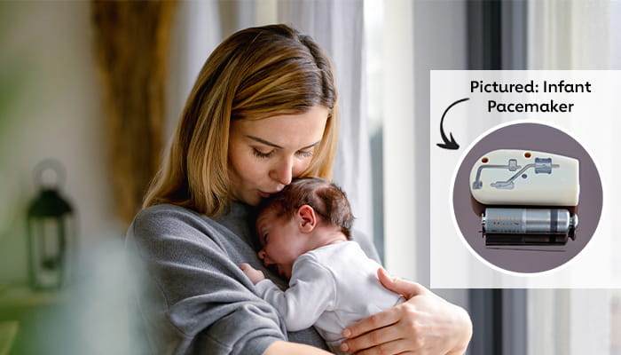
Every saved life begins with research. We’re extending our 4X gift match to help fund lifesaving interventions like the infant pacemaker.
-

CPR, AED or First Aid Training
Be prepared: Learn CPR, AED, and First Aid skills that can save a life.
-

Support Network Community
Join our online community of patients, survivors and caregivers.
-
Professional Membership
Build your professional career and start making an impact today.
-

Volunteer Opportunities
Help us create a healthier world free of heart disease and stroke.
How we are changing the future of health together
Since the American Heart Association’s founding in 1924, deaths from cardiovascular diseases have been cut in half. And yet, there are still so many lives to be saved. By driving breakthroughs in science, policy and care, together we can continue to advance health and transform lives every day.
19 Million+
High blood pressure patients helped
Our high blood pressure quality care initiative ensures lifesaving care for people at risk for heart disease.
Our high blood pressure quality care initiative ensures lifesaving care for people at risk for heart disease.
$6 Billion+
Invested in pioneering research leading to lifesaving breakthroughs
The American Heart Association is the largest non-profit, non-governmental funder of cardiovascular and cerebrovascular research.
The American Heart Association is the largest non-profit, non-governmental funder of cardiovascular and cerebrovascular research.
22 Million
People trained in CPR every year
9 out of 10 people who have a cardiac arrest outside of the hospital die. If performed immediately, CPR can double or triple a person's chances of survival.
9 out of 10 people who have a cardiac arrest outside of the hospital die. If performed immediately, CPR can double or triple a person's chances of survival.

Summer Heat Can Sneak Up Before You Feel Thirsty
Hydration does more than help with thirst. It helps your heart pump blood and your body regulate temperature. Before you spend time on the beach or in your backyard, know the simple signs to watch for and how to stay ahead of the heat.

NEW! 2026 Cardiovascular-Kidney-Metabolic (CKM) Syndrome Guideline
Heart disease goes beyond the heart. Learn about the first ever guideline for managing CKM syndrome including prevention, screening, and care across the overlapping cardiovascular, kidney and metabolic risk factors.

Make the Most out of Telehealth
Telehealth makes it easier to get the care you need for conditions like high blood pressure and diabetes—without leaving home. We have new tips, tools, and trusted resources to help you feel confident about using virtual care.

New American Heart Association president aims to close the gap between discovery and care
Manesh Patel’s signature curiosity and drive fuel his focus on closing the divide between discovery and care.News and Stories
News and Stories from the American Heart Association
Read more News and Stories 

Join us in saving lives
Get updates on groundbreaking research, inspiring survivor stories, and ways you can help save lives—all delivered to your inbox.
Find a Heart Walk in your area
Heart Walk is the American Heart Association's premiere event for raising funds to help save lives from heart disease and stroke.